MEG
Magnetoencephalography (MEG) consists in the study of the magnetic fields associated with physiological and pathological activity in the brain. In fact, all electrical currents - in power lines or brain cells - generate a surrounding magnetic field. The pattern of this magnetic fields can be used to determine the location, orientation and strength of the currents that generates it. The elementary “magnetic field generator” of the brain is the single neuron. When a sufficiently large population of neurons receives synaptic inputs within a short time-window, the dendritic currents will sum up, producing a field which is large enough to be detected outside the head. The neuromagnetic fields are very small, typically in the order of 100 fT. To successfully detect neuromagnetic fields, superconducting devices must be used. These devices, namely SQUIDs (Superconducting QUantum Interference Devices), were introduces in the late 1960s and are the components present in every MEG system. Today's whole-head MEG systems contain a large number of SQUIDs (between 100 to 300) connected to sensor coils in a configuration roughly following the curvature of the head. In addition to a very sensitive device, a successful MEG measurements demands for a magnetically quiet environment. To that, MEG system needs to be placed in a magnetic shielded room. The main advantage of the neuromagnetic method over the more traditional measurement of brain electrical activity, such as electroencephalography (EEG), is mainly due to the "transparency" of biological tissues to magnetic field. This allows a better resolution in identifying the location and strength of the sources responsible for the specific activity under investigation, of course by the use of suitable algorithms for data analysis. The good spatial resolution of the neuromagnetic method therefore permits to obtain a real functional imaging of the brain. This information may be integrated with the anatomical imaging as given by CT, MRI or metabolic imaging as given by PET and functional MRI.
MEG at ITAB
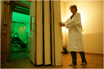
Within ITAB an MEG system have been developed in the frame of an international co-operation among research institutions and companies. The system features 165 sensors, 153 of which displaced on a helmet-like surface. The sensing elements are integrated dc-SQUID magnetometers, featuring a field noise of about 5 fT Hz-1/2. The system is placed inside a five layer magnetic shielded room.