Risorse
Core Facilities
Infrastruttura Informatica del CED ITAB – Istituto Tecnologie Avanzate Biomediche condiviso con Dipartimento DNISC – Neuroscienze, Imaging e Scienze Cliniche
1. Panoramica generale
L'infrastruttura di calcolo dell'ITAB, condivisa con il Dipartimento DNISC, è costituita da un insieme eterogeneo di nodi di calcolo ad alte prestazioni, workstation scientifiche e sistemi di storage centralizzato. La piattaforma è progettata per supportare attività di elaborazione e analisi di dati di neuroimaging nelle modalità EEG, EMG, MEG, fMRI e MRI, con impiego prevalente di software specializzati quali FSL, SPM, FreeSurfer, EEGLAB e Brainstorm.
L'infrastruttura comprende:
- 12 workstation scientifiche ad alte prestazioni;
- 2 server rack dedicati all'elaborazione e alla gestione dei dati;
- 1 sistema di storage centralizzato ad alta capacità.
2. Workstation scientifiche
Le 12 workstation sono sistemi multi-core basati su processori Intel Xeon, dotati di GPU NVIDIA o AMD e configurate con ampie quantità di RAM. Ogni sistema è identificato da un nome mnemonico e un indirizzo IP fisso sulla rete interna.
- Sestieri
- CPU: Intel Xeon E5-2630 – 10 core × 2 socket (20 core totali)
- RAM: 64 GB DDR4
- GPU: NVIDIA GTX 1050 2 GB
- Sestieri/Baldassarre/Committeri
- CPU: Intel Xeon 3204 – 6 core × 1 socket
- RAM: 64 GB DDR4
- GPU: —
- Perrucci
- CPU: Intel Xeon E5-2630 v4 – 10 core × 2 socket (20 core totali)
- RAM: 64 GB DDR4
- GPU: NVIDIA Quadro K1200 4 GB
- Perrucci
- CPU: Intel Xeon Silver 4114 – 10 core × 2 socket (20 core totali)
- RAM: 96 GB DDR4
- GPU: NVIDIA GeForce GTX 1070 Ti 8 GB
- Della Penna (2 sistemi identici)
- CPU: Intel Xeon Silver 4208 – 8 core × 2 socket (16 core totali)
- RAM: 64 GB DDR4
- GPU: NVIDIA Quadro P1000 4 GB
- Wise
- CPU: Intel Xeon 4210 – 10 core × 2 socket (20 core totali)
- RAM: 128 GB DDR4
- GPU: NVIDIA Quadro RTX 4000 8 GB
- Pizzella (A)
- CPU: Intel Xeon w5-2565X – 18 core × 1 socket
- RAM: 128 GB DDR5
- GPU: NVIDIA Quadro T400 4 GB
- Pizzella (B)
- CPU: Intel Xeon 4210R – 10 core × 1 socket
- RAM: 64 GB DDR4
- GPU: NVIDIA Quadro RTX 4000 8 GB
- Del Gratta (A)
- CPU: Intel Xeon E5-2630 v3 – 8 core × 1 socket
- RAM: 32 GB DDR4
- GPU: AMD FirePro W4100
- Del Gratta (B) (2 sistemi identici)
- CPU: Intel Xeon 4210R – 10 core × 1 socket
- RAM: 64 GB DDR4
- GPU: NVIDIA Quadro RTX 5000 16 GB
3. Server di elaborazioni
Il CED dispone di due server rack con configurazioni RAID ridondanti per il disco di sistema e per i volumi dati, garantendo continuità operativa in caso di guasto hardware.
- DELL PowerEdge R740XD – Server Rack 2U (nodo principale)
- CPU: Intel Xeon Gold 6238R – 28 core × 2 socket (56 core / 112 thread totali)
- RAM: 256 GB DDR4
- Storage OS: 1 TB SSD in RAID 1
- Storage dati: 27 TB SAS in RAID 6
- Lenovo ThinkSystem SR590 – Server Rack 1U (nodo secondario)
- CPU: Intel Xeon Silver 4208 – 8 core × 2 socket (16 core / 32 thread totali)
- RAM: 128 GB DDR4
- Storage OS: 300 GB SAS in RAID 1
- Storage dati: 60 TB SAS in RAID 6
4. Sistema di storage centralizzato
Lo storage centralizzato è realizzato tramite un array DELL PowerVault ME4024 espanso con unità aggiuntiva ME412, per una capacità complessiva di circa 116 TB lordi. La configurazione RAID 6 garantisce la tolleranza al guasto contemporaneo di due dischi, assicurando la protezione dei dataset di ricerca non riproducibili.
- DELL PowerVault ME4024 + ME412
- Volume HDD: 100 TB in RAID 6 – archiviazione a lungo termine di dati grezzi e processati;
- Volume SAS: 16 TB in RAID 6 – accessi ad alte prestazioni per pipeline di analisi attive.
5. Riepilogo risorse di calcolo
- Workstation scientifiche: 14 unità
- Server rack: 2 unità (DELL R740XD + Lenovo SR590)
- Core CPU totali – Workstation: 166 core fisici / 332 thread
- Core CPU totali – Server: 72 core fisici / 144 thread
- RAM totale – Workstation: circa 896 GB
- RAM totale – Server: 384 GB (256 GB + 128 GB)
- Storage centralizzato: circa 116 TB lordi in RAID 6 (DELL ME4024 + ME412)
- Storage interno server: circa 87 TB lordi in RAID 6 (27 TB R740XD + 60 TB SR590)
6. Destinazione d'uso e ambiti applicativi
L'intera infrastruttura è dimensionata per supportare le esigenze computazionali tipiche della ricerca in neuroimaging clinico e cognitivo. Gli ambiti di utilizzo principali includono:
- Elaborazione e analisi di immagini MRI strutturale e funzionale (fMRI), con pipeline FSL, SPM, FreeSurfer;
- Analisi di segnali elettrofisiologici (EEG, EMG) e magnetoencefalografici (MEG) con toolbox EEGLAB, FieldTrip, Brainstorm;
- Archiviazione sicura e ridondante dei dataset di imaging clinico e sperimentale a lungo termine;
- Supporto a pipeline di machine learning e deep learning su dati neuroimaging mediante acceleratori GPU NVIDIA;
- Condivisione delle risorse di calcolo tra i gruppi di ricerca ITAB e DNISC per attività collaborative e progetti di ricerca finanziati.
LA TECNICA
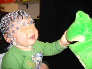
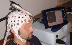
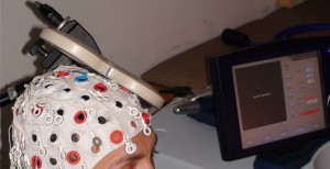
L'elettroencefalografia (EEG) è la registrazione dell'attività elettrica lungo il cuoio capelluto, prodotta dall'attivazione dei neuroni nel cervello. In ambito clinico, l'EEG si riferisce alla registrazione dell'attività elettrica spontanea del cervello in un breve periodo di tempo, solitamente 20-40 minuti, rilevata da più elettrodi posizionati sul cuoio capelluto. In neurologia, la principale applicazione diagnostica dell'EEG è nel caso dell'epilessia, poiché l'attività epilettica può generare anomalie evidenti in un esame EEG standard. Tra i derivati della tecnica EEG figurano i potenziali evocati (PE), che comportano la media dell'attività EEG sincronizzata con la presentazione di uno stimolo di qualche tipo (visivo, somatosensoriale o uditivo). I potenziali evento-correlati si riferiscono a risposte EEG medie sincronizzate con l'elaborazione più complessa degli stimoli; questa tecnica è utilizzata nelle scienze cognitive, nella psicologia cognitiva e nella ricerca psicofisiologica.
STRUMENTAZIONI
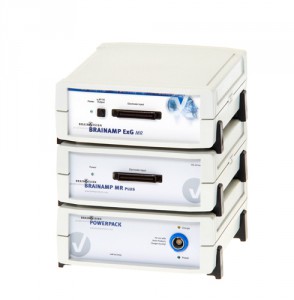
EEG BrainAmp (Brain Products)
2x BrainAmp MR plus (32 canali per unità)
1x BrainAmp MR (32 canali)
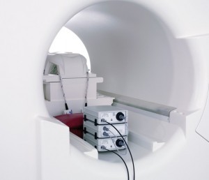
SyncBox per registrazioni simultanee EEG e fMRI
BrainAmp ExG MR (8 canali)
Sensori MRI: Brain Products lancia il tanto atteso GSR (Galvanic Skin Response)
2x BrainCap MR 64 canali
2x PowerPack (batteria ricaricabile compatibile con la risonanza magnetica)
BrainVision Recorder (software di registrazione)
BrainVision RecView (software per l'analisi dei dati in tempo reale)
BrainVision Analyzer 2 (software di analisi)
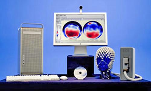
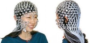
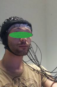
EEG Geodesic (GES 300)
- HydroCel Geodesic Sensor Net (HCGSN) 130 con 128 canali (disponibile per bambini e adulti)
- Mac Pro + MacBookPro come computer di acquisizione
- Desktop + Laptop per la presentazione degli stimoli con software E-Prime
- Amplificatore Net Amps 300
- Software Net Station per l'acquisizione EEG ed elaborazione
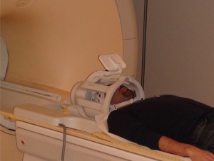
La risonanza magnetica funzionale (fMRI) è una tecnica di neuroimaging non invasiva che rileva i cambiamenti nell'attività neuronale
SISTEMI DISPONIBILI
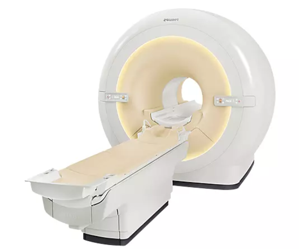
MR2 - Philips 3T SmartPath to dStream
- Bobina dS Head 32 canali - 3.0T
- BOLD Specialist
- FiberTrak Specialist
- 3D ASL Neuro Specialist
- MultiBand SENSE
- Spectroscopy Specialist
- NeuroScience Specialist
- Acquisizione Fisiologica Wireless PPU
- Sistema Proiettore EIKI LC XG-250L con lente aggiuntiva
- Sistema Audio NordicNeuroLab con Cuffie e Console di Comunicazione
- BrainVision BrainAmp MR plus 64 canali (amplificatori EEG per acquisizione combinata EEG & fMRI)
- Sistema controller Lumina LP-400 con tastierini di risposta a Fibra Ottica
- Due Stimolatori Elettrici Digitimer modello DS7A
- Computer desktop per la registrazione dei dati EEG
- Computer desktop per la presentazione di stimoli
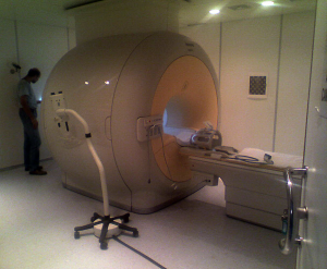
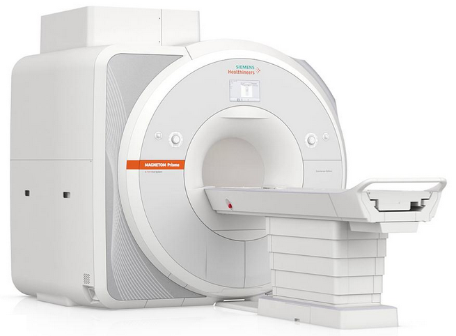
MR3 - MAGNETOM Prisma 3T
- Head Neck 20
- Head Neck 64
- Head 32
L'imaging a infrarossi (IR) è una tecnica che permette la misurazione dell'emissione infrarossa termica del corpo umano.
L'imaging a infrarossi medicale (conosciuta anche come termografia medicale) ha avuto inizio negli anni '70.
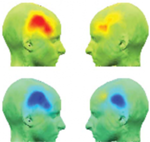
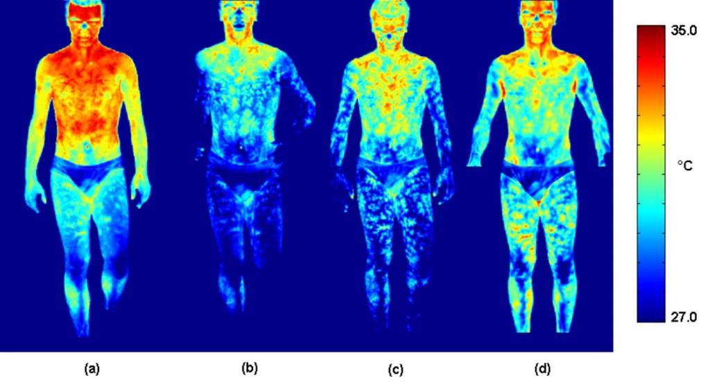 Immagini termiche a infrarossi: lo schema a falsi colori mostra la modifica della distribuzione della temperatura cutanea durante un esercizio atletico.
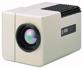
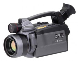
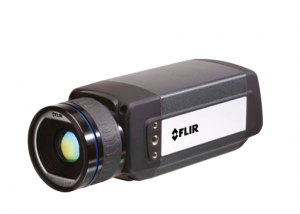
- Thermal IR Camera FLIR SC3000, 320x200, QWIP, NETD: 20 mK @ 30°C
- Thermal IR Camera FLIR SC660, 640x480, Microbolometer, NETD: 30 mK @ 30°C
- Thermal IR Camera FLIR A655sc, 640x480, Microbolometer, NETD: 30 mK @ 30°C (una usata per l'insegnamento e la formazione)
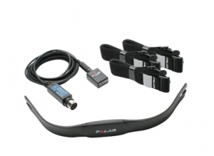
- Sistema Full PowerLab ADInstruments
- Wireless Detectors per Psicofisica
- Computers per stimolazione triggerata
- Software per la preparazione e la somministrazione di stimoli
LA TECNICA
Le misurazioni di Near Infra-Red Spectroscopy (NIRS) si basano sulla valutazione delle proprietà ottiche dei tessuti e della loro dipendenza da parametri fisiologici connessi all'attività cerebrale corticale. Nel range spettrale tra 600nm e 900nm, ossiemoglobina e deossiemoglobina sono le principali fonti responsabili dell'assorbimento della radiazione. Pertanto, è possibile valutare le proprietà ottiche del tessuto e stimare la concentrazione di questi due cromofori effettuando misurazioni in questo intervallo di lunghezze d'onda. Negli ultimi dieci anni, NIRS è diventato uno strumento utile per condurre studi di neuroimaging.
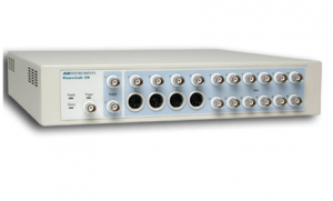
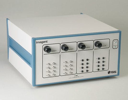
STRUMENTI DISPONIBILI
Un sistema NIRS Imagent ISS a 32 canali per il neuro-imaging verrà installato a breve nel laboratorio e utilizzato per studiare lo sviluppo delle funzioni cerebrali nei neonati.
STRUMENTI DISPONIBILI
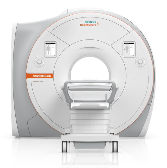
MR1 - Siemens MAGNETOM SOLA 1.5T (Datasheet)
- Bobina mammella predisposta per biopsia
- Bobina mammella per imaging
- Pacchetto encefalo
- Pacchetto Cardio
- Consolle di post-elaborazione
- Bobina copertura arti inferiori
MR2 - Philips 3T SmartPath to dStream
- Bobina dS Torso
- Bobina dS Flex-M
- Bobina dS Head 32 canali - 3.0T
- ScanTools Pro
- 3D SpineVIEW
- MultiVaneXD
- BOLD Specialist
- FiberTrak Specialist
- SWI Specialist
- 3D ASL Neuro Specialist
- MultiBand SENSE
- Spectroscopy Specialist
- mDIXON XD TSE-FFE Specialist
- mDIXON Body Fat Quant Specialist
- Cardiac Expert Specialist
- 4D-TRAK XD
- Cardiac Quant
- Coronary Acquisition
- 3D PelvisVIEW
- lmaging ZOOM Diffusion
- Whole Body Specialist
MR3 - MAGNETOM Prisma 3T
- Head Neck 20
- Head Neck 64
- Head 32
- Flex Small 4
- Body 18
- Spine 32
- Body 18
LA TECNICA
La Magnetoencefalografia (MEG) consiste nello studio dei campi magnetici associati all'attività fisiologica e patologica del cervello. Infatti, tutte le correnti elettriche – sia quelle nelle linee elettriche che quelle nelle cellule cerebrali – generano un campo magnetico circostante. Il pattern di questi campi magnetici può essere usato per determinare la posizione, l'orientamento e la forza delle correnti che lo generano. Il "generatore di campo magnetico" elementare del cervello è il singolo neurone. Quando una popolazione sufficientemente ampia di neuroni riceve input sinaptici entro un breve intervallo di tempo, le correnti dendritiche si sommano, producendo un campo sufficientemente grande da essere rilevato all'esterno della testa. I campi neuromagnetici sono molto piccoli, tipicamente dell'ordine di 100 fT. Per rilevare con successo i campi neuromagnetici, devono essere utilizzati dispositivi superconduttori. Questi dispositivi, chiamati SQUID (Superconducting QUantum Interference Devices), furono introdotti alla fine degli anni '60 e sono i componenti presenti in ogni sistema MEG. Gli attuali sistemi MEG a testa intera contengono un gran numero di SQUID (tra 100 e 300) collegati a bobine sensore in una configurazione che segue approssimativamente la curvatura della testa. Oltre a un dispositivo molto sensibile, una misurazione MEG di successo richiede un ambiente magneticamente silenzioso. Per questo, il sistema MEG deve essere collocato in una stanza schermata magneticamente. Il principale vantaggio del metodo neuromagnetico rispetto alla misurazione più tradizionale dell'attività elettrica cerebrale, come l'elettroencefalografia (EEG), è dovuto principalmente alla "trasparenza" dei tessuti biologici al campo magnetico. Questo permette una migliore risoluzione nell'identificare la posizione e la forza delle sorgenti responsabili della specifica attività in esame, ovviamente attraverso l'uso di algoritmi adatti per l'analisi dei dati. La buona risoluzione spaziale del metodo neuromagnetico consente quindi di ottenere un vera e propria imaging funzionale del cervello. Queste informazioni possono essere integrate con l'imaging anatomico fornito da TC, risonanza magnetica o imaging metabolico fornito da PET e risonanza magnetica funzionale.
MEG ALL'ITAB
All'interno dell'ITAB è stato sviluppato un sistema MEG nell'ambito di una cooperazione internazionale tra istituti di ricerca e aziende. Il sistema dispone di 165 sensori, di cui 153 disposti su una superficie a forma di casco. Gli elementi sensibili sono magnetometri SQUID DC integrati, con un rumore di campo di circa 5 fT Hz-1/2. Il sistema è posizionato all'interno di una stanza schermata magneticamente a cinque strati.
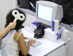
La struttura di Psicofisica permette l'implementazione di esperimenti comportamentali su soggetti normali e pazienti neuropsicologici, il testing preliminare di stimoli e procedure da effettuare successivamente con altre strutture principali (es. fMRI, MEG, ecc.) così come l'addestramento di soggetti/pazienti sperimentali.
La struttura è equipaggiata con piattaforme hardware-software che consentono l'erogazione di stimolazione visiva e uditiva, la registrazione delle risposte del soggetto con diversi effettori (risposte manuali, podaliche e vocali) e il tracciamento del movimento 3D di segmenti corporei (tramite un dispositivo elettromagnetico: 3 Space Fastrak, Polhemus Navigation; Colchester, VT, USA).
I movimenti oculari possono anche essere tracciati, tramite un eye tracker a infrarossi monoculare remoto (ISCAN ETL 400; RK-826PCI), che utilizza un metodo di riflessione pupilla scura-cornea basato su video per registrare le posizioni degli occhi durante l'analisi di immagini e scene, oltre a verificare semplicemente il mantenimento della fissazione. Con una frequenza di campionamento di 120 Hz, consente una registrazione ad alta risoluzione della posizione dell'occhio e della dimensione della pupilla in tempo reale con una precisione tipicamente migliore di 0,3° su un intervallo orizzontale e verticale di +/- 20°. Un pannello in plexiglass con LED montati è disponibile per lo studio dei movimenti di raggiungimento verso bersagli visivi nello spazio.
Gli stimoli vengono solitamente generati da un computer di controllo situato all'esterno della stanza di psicofisica, che esegue il software personalizzato GagLab (sviluppato da Gaspare Galati presso il Dipartimento di Psicologia, Sapienza Università di Roma, Italia), implementato in MATLAB (The MathWorks Inc., Natick, MA, USA) utilizzando Cogent 2000 (sviluppato presso FIL e ICN, UCL, Londra, UK) e Cogent Graphics (sviluppato da John Romaya presso il LON, Wellcome Department of Imaging Neuroscience, UCL, Londra, UK), e che consente la presentazione di stimoli visivi e uditivi sincronizzata con precisione al millisecondo, sincronizzata con fMRI, TMS, EEG, MEG. Vengono utilizzati anche altri software, come E-Prime.
È inoltre disponibile una simulazione in legno del bore (apertura) del magnete fMRI per riprodurre l'impostazione fMRI in un modo il più possibile simile a quello che i soggetti sperimenteranno (cioè, sdraiati su un semicerchio stretto).
PICTURE LEGENDS:
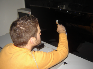
- Impostazione per un tipico esperimento che coinvolge TMS, eye tracking, stimolazione visiva e pressione dei pulsanti
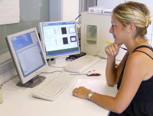
- Le postazioni di lavoro degli sperimentatori consentono il controllo in tempo reale del comportamento e dei movimenti oculari del soggetto
- Setup per lo studio dei movimenti di avvicinamento verso LED montati su pannello in plexiglass
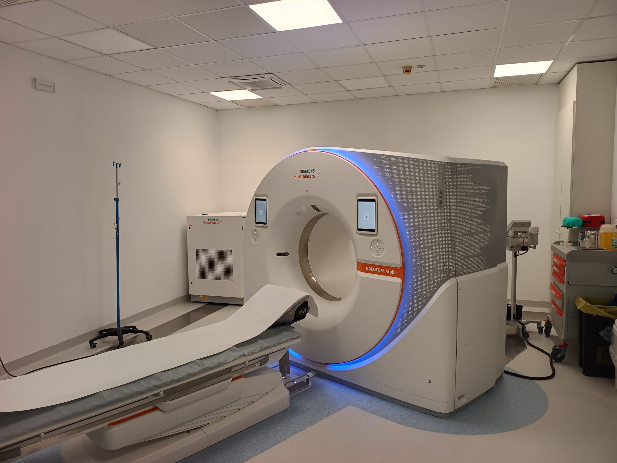
NAEOTOM Alpha Dual photon-counting
Il cuore di NAEOTOM Alpha è un detettore, radicalmente nuovo, per il conteggio dei fotoni. Il detettore QuantaMax converte direttamente i raggi X in un segnale elettrico, che viene poi utilizzato per creare un'immagine. L'energia di ogni raggio X viene misurata, quindi le informazioni spettrali sono disponibili per ogni scansione e le immagini sono ad alto contrasto con un'elevata risoluzione spaziale a parità di dose. La combinazione dell'alta risoluzione spaziale del detettore QuantaMax della Tac Photon Counting con la risoluzione temporale della nostra Dual Source consente la visualizzazione di dettagli estremamente precisi per una maggiore affidabilità diagnostica.
LA TECNICA
La stimolazione magnetica transcranica (TMS) è un metodo non invasivo per eccitare i neuroni nel cervello: deboli correnti elettriche vengono indotte nel tessuto tramite campi magnetici che cambiano rapidamente (induzione elettromagnetica). La TMS può essere utilizzata per integrare altri metodi delle neuroscienze e fornisce una metodologia unica per determinare il vero significato funzionale dei risultati degli studi di neuroimaging e la relazione causale tra l'attività cerebrale focale e il comportamento.
STRUMENTI DISPONIBILI
- TMS Magstim Super Rapid²
- TMS Magstim BiStim²
- Due Double 70mm Alpha Coil
- Single 90mm Remote Control Coil
- D360 8-Channel Patient Amplifier (per la registrazione di Elettroencefalografia, Elettromiografia o Potenziali Evocati)
- CED Power 1401 (interfaccia ad alte prestazioni per acquisizione e analisi dati)
- Polhemus Fastrack Neuronavigation System (Quattro Short Ranger TX1 + 8" Stylus)
- SofTAX Software for the neuronavigation (permette la neuronavigazione sul cervello del soggetto anche senza MPRAGE).
- Mechanical arm (per mantenere l'impugnatura della bobina angolata nella posizione corretta)
- Computer desktop per la presentazione di stimoli visivi
Scopri cosa vuol dire essere dell'Ud'A
SEDE DI CHIETI
Via dei Vestini,31
Centralino 0871.3551
SEDE DI PESCARA
Viale Pindaro,42
Centralino 085.45371
email: info@unich.it
PEC: ateneo@pec.unich.it
Partita IVA 01335970693


